Adelaide & Hills HAND THERAPY
|
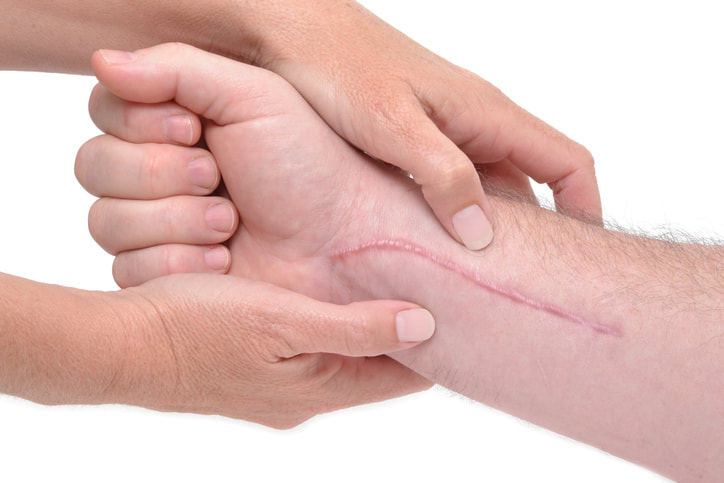
We all have a scar somewhere on our body.
Scarring is a natural process that the body undergoes when healing from a wound. On the skin, scar tissue is mostly made of collagen. The collagen is a protein which is produced to close the edges of a wound to protect us from infection. Hypertrophic and keloid scar tissue sometimes forms when wound healing is delayed and / or excessive collagen is produced. This more likely occurs when there is damage to the deeper layers of skin or following an infection. The excess collagen production results in a scar becoming raised, thick and often darker than the surrounding skin. These scars are not dangerous, however can cause discomfort and limit movement. And for some people had a negative psychological impact. Hypertrophic scar Hypertrophic scars are thick, hard, raised, can be smooth or rough and are frequently darker than the surrounding skin (pink to deep purple). They often occur following a deep cut (laceration), surgical scar or burn. Hypertrophic scars are most likely to form over mobile joints where the skin is required to stretch and move as the joint below changes position. Keloid scar A keloid scar differs from other scars in that it exceeds the border of the original wound. That is, it grows larger than the initial area of injury. These scars are always dark, raised and thick, they can be rubbery, shiny and may be itchy and are sometimes even painful. Keloid scars often result from the site of a pimple, insect bite or piercing. The good news is that there are strategies to help manage the way a scar grows and matures. Seeking guidance from a professional who has experience in burns & scar management will get you set with what you can do to help minimise the appearance of your scar.
0 Comments
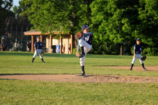
We know that elbow pain can result from common causes such as a broken bone, tendinitis and arthritis. But when a child or teen report having a sore elbow after sport, without incidence of sudden trauma - the cause may not seem so clear. It is worth investigating whether they have developed Osteochontritis dissecans.
What is Osteochondritis Dissecans? Elbow Osteochondritis dissecans is a condition at the elbow joint, where bone under the cartilage dies due to lack of blood flow. The bone & cartilage become damaged which results in pain and decreased movement at the joint. This may result sudden injury and fracture (like a fall on the outstretched hand). Or from damage to the bone from repetitive trauma (for example during participation in sporting activities particularly involving throwing actions). Symptoms of Osteochondritis dissecans include:
Diagnosis of Osteochondritis dissecans: If you think you or your child may have developed Osteochondritis dissecans it is best to book an appointment to discuss with your doctor. As diagnosis is usually made from reviewing x-ray or MRI of the involved elbow. Treatment for Osteochondritis dissecans: Treatment is determined by the degree of damage to the bone. In some instances, symptoms can be managed without operation with hand therapy alone. However often surgical advice is recommended, as an operation may be required to remove any bone fragment from the joint. |
Author Jo MarshClick here to edit Archives
March 2020
|


 RSS Feed


